
Knee Pain: Is It Your ACL, MCL, or Meniscus?
Twisted your knee and not sure what you hurt? Here's how the ACL, MCL, and meniscus differ, the simple clinical tests that point to each one, and when to get checked.
Knee pain after a twist, pivot, or awkward landing is one of the most common things we see. Three structures get hurt more than any others: the ACL, the MCL, and the meniscus. Each has its own anatomy, its own way of getting injured, and its own set of clinical tests that help point to the cause.
This guide breaks them down. It's meant to help you understand your knee — not to replace an exam. If your knee is swollen, locking, or giving way, get it evaluated.
A quick tour of the knee
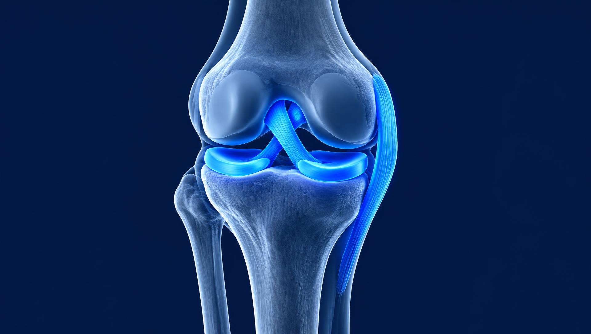
The knee is where your thigh bone (femur) meets your shin bone (tibia). Because the bones themselves aren't very stable, the knee relies on soft tissue:
- Ligaments connect bone to bone and stop unwanted motion.
- The menisci are two C-shaped pads of cartilage that cushion and stabilize the joint.
- Muscles (quads, hamstrings, calves, hips) control the knee during movement.
The ACL (Anterior Cruciate Ligament)
Anatomy
The ACL runs diagonally through the middle of the knee, from the back of the femur to the front of the tibia. Its main job is to stop the shin from sliding forward and to control rotation.
How it gets hurt
Most ACL tears are non-contact: planting and cutting, pivoting, or landing from a jump with the knee caving inward. People often describe:
- A "pop" at the time of injury
- Fast swelling within a few hours
- The knee feeling unstable or "giving way"
Tests that point to the ACL
- Lachman test — With the knee bent about 20–30°, the examiner stabilizes the thigh and pulls the shin forward. Extra movement or a soft "end feel" suggests an ACL tear. Research reviews consistently rate this as the most accurate single clinical test for the ACL.
- Anterior drawer test — Lying on your back with the knee bent to 90° and the foot flat, the examiner pulls the top of the shin forward. More forward glide than the other side suggests an ACL injury. It's more reliable in older (chronic) injuries than right after an injury, when swelling and guarding can hide laxity.
- Pivot shift test — The examiner moves the knee from straight to bent while applying a rotational and inward force. A "clunk" as the joint shifts is highly specific for an ACL tear, but it's hard to do on a painful, guarded knee.
The MCL (Medial Collateral Ligament)
Anatomy
The MCL runs along the inside of the knee, from the femur to the tibia. It resists valgus force — the knee being pushed inward toward the other knee.
How it gets hurt
Classic MCL injuries come from a blow to the outside of the knee (a tackle or collision) or a twist where the knee collapses inward. Signs include:
- Pain and tenderness along the inside of the knee
- Swelling that's often more localized than with an ACL tear
- Pain with side-to-side cutting
MCL injuries are graded 1 (stretched) to 3 (fully torn). Most heal well without surgery.
Tests that point to the MCL
- Valgus stress test — The examiner pushes the knee inward while stabilizing the ankle, first at about 30° of bend and then fully straight. Pain or gapping at 30° points to the MCL. Gapping when fully straight suggests a more serious injury that may also involve the ACL or other structures.
- Varus stress test — The opposite force, pushing the knee outward. This checks the LCL on the outside of the knee and helps rule other ligaments in or out when comparing sides.
- Palpation — Tenderness right along the MCL, especially near where it attaches to the femur, is a useful clue.
The Meniscus
Anatomy
Each knee has two menisci — medial (inside) and lateral (outside) — sitting between the femur and tibia. They absorb shock, spread out load, and help keep the joint stable.
How it gets hurt
- In younger, active people: a twist on a bent, weight-bearing knee (squatting and turning, pivoting in sports).
- In people over 40: degenerative tears can happen with very little trauma — sometimes just getting up from a squat.
Common signs:
- Pain along the joint line (inside or outside of the knee)
- Slower swelling, often over 24–48 hours
- Catching, clicking, or locking
- Pain with deep squatting or twisting
Tests that point to the meniscus
- Thessaly test — Standing on the injured leg with the knee bent about 20° (holding the examiner's hands for balance), you rotate your body and knee in and out three times. Pain at the joint line or a catching sensation suggests a meniscus tear. The original study reported high accuracy, though later research found it performs best when combined with other tests rather than used alone.
- McMurray test — Lying down, the examiner bends and rotates the knee while straightening it. A painful click along the joint line suggests a tear.
- Joint line tenderness — Pressing along the gap between the femur and tibia. Tenderness here is a common (though not perfectly specific) sign.
Putting it together
| ACL | MCL | Meniscus | |
|---|---|---|---|
| Typical injury | Non-contact pivot or landing | Blow or force to outside of knee | Twist on a bent knee, or degeneration |
| Pop? | Often | Sometimes | Rarely |
| Swelling | Fast (hours) | Mild, localized | Slow (1–2 days) |
| Where it hurts | Deep in the knee | Inside of the knee | Joint line |
| Key feeling | Giving way | Pain with side-to-side load | Catching or locking |
| Key tests | Lachman, anterior drawer, pivot shift | Valgus stress at 30° and 0° | Thessaly, McMurray, joint line tenderness |
Important: these injuries often happen together. A common combination is ACL + MCL + medial meniscus. No single test is perfect — research shows clinical tests are most accurate when a trained provider combines your history, several tests, and imaging when needed.
When to get it checked right away
- The knee locks and you can't straighten it
- You can't put weight on it
- Rapid, significant swelling after a pop
- The knee repeatedly gives way
- Numbness, color change, or severe deformity (go to urgent care or the ER)
How we help at Indy Spine and Rehab
At our Carmel and Delphi clinics, Dr. Owen and our team:
- Take a detailed history of how the injury happened.
- Perform a full knee exam, including the ligament and meniscus tests above.
- Refer for imaging (like an MRI) or to an orthopedic specialist when it's needed.
- Build a rehab plan — soft tissue work, mobility, and progressive strength for the quads, hamstrings, and hips to restore stability and get you back to activity.
Not sure what's going on with your knee? Book an evaluation and let's find out.
Evidence referenced: Benjaminse et al., 2006 (meta-analysis of ACL clinical tests); Kastelein et al., 2009 (diagnostic value of history and physical exam for knee ligament injuries); Karachalios et al., 2005 (Thessaly test); Goossens et al., 2015 (Thessaly and McMurray accuracy); Hegedus et al., 2007 (systematic review of meniscal tests).