Osteoarthritis: What Happens When You Stop Moving vs. When You Stay Active

The old advice was to rest a painful joint. The evidence now says the opposite — motion is one of the most powerful treatments we have for osteoarthritis. Here is what the research shows.
For decades, people with osteoarthritis (OA) were told the same thing: your joints are "worn out," so stop using them. We now know that advice was backwards. Cartilage, muscle, and the joint itself all respond to load — and when load disappears, the joint gets worse, faster.
Here is what actually happens in the two scenarios, side by side, and what the peer-reviewed literature says about each.
What osteoarthritis actually is
OA is not simple "wear and tear." It is a whole-joint disease involving cartilage breakdown, subchondral bone changes, low-grade synovial inflammation, and progressive muscle weakness around the joint (Hunter & Bierma-Zeisinger, The Lancet, 2019). That last part matters: the muscles around an arthritic joint are part of the disease process, and muscles are the one part you can directly rebuild.
What happens when a patient stops moving
When activity drops to near zero — bed rest, prolonged sitting, "protecting" a sore knee by not using it — several things happen quickly:
- Cartilage thins and softens. Cartilage has no blood supply. It gets nutrients from joint fluid that only circulates when the joint is loaded and unloaded through movement. Immobilization studies show measurable cartilage thinning within weeks, and the changes track closely with loss of loading (Vanwanseele et al., Arthritis & Rheumatism, 2002).
- Muscle atrophies fast. Quadriceps strength — the single best mechanical protector of the knee — can drop 8–17% in the first week of immobilization or bed rest (Wall et al., Acta Physiologica, 2013). Weak quads are independently associated with knee OA progression and pain (Segal & Glass, Physician and Sportsmedicine, 2011).
- Systemic inflammation goes up. Sedentary behavior raises circulating inflammatory markers like IL-6 and CRP, which are the same signals that drive synovitis inside the joint (Yates et al., American Journal of Preventive Medicine, 2012).
- Pain sensitivity increases. Physical inactivity is linked to central sensitization and higher pain scores in people with OA, independent of imaging findings (Fingleton et al., Osteoarthritis and Cartilage, 2015).
- Function and independence drop. Long-term inactivity in adults with OA predicts faster progression to disability and higher rates of joint replacement (Dunlop et al., BMJ, 2014).

The short version: the joint you are trying to protect by resting is exactly the joint that gets worse from resting.
What happens when a patient stays active
The evidence for the other direction is unusually strong for musculoskeletal medicine. Both the American College of Rheumatology and OARSI now list exercise as a core, first-line treatment for hip and knee OA — ahead of injections and imaging (Kolasinski et al., Arthritis Care & Research, 2020 (ACR guideline); Bannuru et al., Osteoarthritis and Cartilage, 2019 (OARSI guideline)).
What the trials show when patients keep moving:
- Pain and function improve. A Cochrane review of 54 randomized trials with over 3,900 knee OA patients found that land-based exercise produces moderate reductions in pain and improvements in function that persist for months after the program ends (Fransen et al., Cochrane Database of Systematic Reviews, 2015).
- Effect sizes rival medication. A network meta-analysis found exercise therapy produced pain relief comparable to NSAIDs — without the GI, cardiovascular, or renal risks (Weng et al., British Journal of Sports Medicine, 2023).
- Cartilage responds to loading. Moderate exercise in knee OA patients is associated with improved cartilage glycosaminoglycan content on MRI — the opposite of what happens with disuse (Roos & Dahlberg, Arthritis & Rheumatism, 2005).
- Fewer joint replacements. A long-term follow-up of the BOA program in Sweden — a supervised education-and-exercise protocol — showed roughly a 44% lower rate of knee replacement surgery at 5 years compared to matched controls (Dell'Isola et al., Osteoarthritis and Cartilage, 2020).
- Even walking helps. In adults over 50 with knee OA, walking for exercise was associated with less frequent new knee pain and less medial cartilage loss over 4 years (Lo et al., Arthritis & Rheumatology, 2022).
Why "stay active" beats "rest it"
Three mechanisms explain the gap:
- Load is nutrition for cartilage. Cyclic loading pumps synovial fluid in and out of the cartilage matrix. No load, no nutrition.
- Strong muscles offload the joint. A stronger quadriceps and hip complex reduces peak contact forces at the knee during every step you take.
- Exercise is anti-inflammatory. Regular activity lowers systemic IL-6 and TNF-α and shifts the local joint environment away from a degenerative pattern (Gleeson et al., Nature Reviews Immunology, 2011).
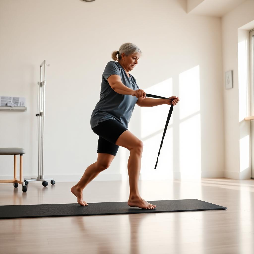
What "active" should look like if you have OA
You do not need to run marathons. The dose in most successful trials is modest and boring — which is a feature, not a bug:
- Aerobic: 30 minutes of walking, cycling, or pool work most days of the week.
- Strength: 2–3 sessions per week targeting the muscles around the affected joint (quads, glutes, calves for the knee and hip).
- Range of motion: Daily gentle movement through full available range — even on flare days, keep the joint moving in a pain-tolerable range.
- Progress gradually. Some soreness during and after exercise is expected and does not mean damage. As a rule of thumb, if pain settles back to baseline within 24 hours, the load was appropriate.
The takeaway
Doing nothing is not a neutral choice with osteoarthritis. It is an active decision that accelerates cartilage loss, muscle atrophy, inflammation, and disability. Consistent, appropriately loaded movement is — by a wide margin — the most evidence-backed, lowest-risk treatment we have.
If your joint hurts and you have been told to "just rest," get a second opinion. If you are not sure what movement is safe for your specific joint, that is exactly what we help patients figure out at Indy Spine and Rehab — we build a loading plan that respects the diagnosis and gets you moving again.
— Dr. Owen Friest, D.C.